Thank you to everyone who joined us for April’s MCOS with Dr. Prashin Unadkat! Missed it? The recording is available here.
Date: May 22nd, 2026
Time: 13:00 UTC
Registration: Please register here.
Title: Deciphering the biological correlate of metabolic connectivity in neurodegenerative diseases

Speaker: Dr. Johannes Gnörich is a clinician scientist at the Department of Nuclear Medicine, LMU University Hospital, LMU Munich, Germany. His research focuses on PET imaging, neuroinflammation, and biomarker development in neurodegenerative diseases. Since his doctoral thesis, he has published several translational studies, including methodological evaluations of single-cell radiotracer detection after in vivo tracer injection (scRadiotracing), aiming to disentangle the cellular sources of PET signals in different inflammatory disorders, including neurodegeneration. These studies contributed to the validation and establishment of novel PET tracers in the field. In addition, Dr. Gnörich integrates advanced network-based imaging analyses, particularly metabolic connectivity approaches, to characterize system-level alterations in brain diseases. Moreover, Dr. Gnörich has a strong record of coordinating academic research projects, including recent multi-center studies. He is actively engaged in international collaborations, serves as a peer reviewer for leading scientific journals, and mentors early-career researchers.
Abstract: Metabolic connectivity derived from [18F]FDG PET is an emerging network-based biomarker in neurodegenerative diseases, yet its underlying biological basis remains insufficiently understood. In this talk, I will present our recent published data combining [18F]FDG-PET with scRadiotracing, demonstrating that metabolic connectivity is strongly influenced by cell-specific glucose uptake patterns of neurons, astrocytes, and microglia. Our findings identify astroglial metabolism as a key driver of age-related network reorganization and show that microglial presence and activation states substantially modulate cerebral metabolic networks in translational mouse models. In addition, I will present our ongoing research and preliminary data using single-cell transcriptomics to further dissect the molecular and subcellular mechanisms underlying metabolic connectivity. Together, these approaches aim to establish metabolic connectivity as a biologically informed imaging marker for neurodegenerative disease.
The MCOS promotes rigor in research and resource sharing. We aim to hold MCOS every third Friday of the month, subject to change due to speaker availability. Please stay tuned for MCOS updates and reminders on social media! Thank you!
Each month, we will feature a member of the MCWG and have a brief Q&A!
This month please enjoy our highlight of Prof Dr Christian Habeck, member of the MCWG Steering Committee.

Prof Dr Christian G Habeck studied Physics and after 3 years of undergraduate study at Siegen University in Germany, went to the UK. He did a Master’s Degree in Elementary Particle Theory at the University of Durham, UK, and finally ended up in a DPhil program at the University of Sussex and the Institut Laue Langevin (ILL, Grenoble), studying neutron decay. The Cold War was barely over.
He realized compared to others he saw at ILL, he was not a gifted experimentalist, and at that time the Life Sciences were a promising new venue for Physicists, so he migrated over. He did a Postdoc in Computational Neuroscience at the Neurosciences Institute in San Diego (now closed), before joining Columbia University.
Prof Dr Christian Habeck has graciously responded to our feature questionnaire:
What sparked your interest in molecular imaging or led you to focus on research in molecular imaging?
Arguably, I have not done much molecular imaging at all.![]() But I like being near people who do it for real! I have dealt with 15O and FDG PET data in the olden days… Recently, I have played with the Monash f-PET data set.
But I like being near people who do it for real! I have dealt with 15O and FDG PET data in the olden days… Recently, I have played with the Monash f-PET data set.
What is your role in the Molecular Connectivity Working Group, and what have you been contributing to or working on within the group?
I see myself in contributing to didactic and methodological debates about connectivity analysis, considering statistical robustness and out-of-sample replication; there are lot of people in MCWG with intricate domain knowledge that I don’t have, but what I do have is a lot of experience of playing with fMRI (and formerly PET) data for group-level covariance analysis.
In what ways do you imagine molecular connectivity will advance our understanding of brain function?
Let me phrase it in terms of the differential impact when comparing MC to resting-state functional connectivity (rs-FC): because of better biological grounding with specific tracers or interventions, I think MC can provide more biological constraints than rs-FC and avoid the tendencies of proliferating levels of analytic complexity. I think the achievements of MCWG to date speak for themselves.
What do you think are the most important challenges in current brain connectivity research, or which unsolved/underappreciated issues should the community address?
I think there are some challenges, specifically: temptations we should try to avoid.
(1) Neuroimaging analytics has become overly derivative, a kind of liberal arts with computers, mainly for intellectual ownership. It’s impossible for one person to have their finger on the pulse of a whole field’s Zeitgeist, and these tendencies are fortunately waning it seems, but in my anecdotal experience as a grant reviewer I have seen a lot of complicated Deep-Learning and AI analytics without any grounding in mechanisms, or even specific ideas about which modalities are important etc. Apart from the fact that neuroimaging science does not ONLY consist of outcome prediction and concerns the discovery of mechanisms in large part too, even on the narrow brief of outcome prediction, the superiority of these techniques is often taken as a given, judging from the absence of validation plans in comparison to simple models using first-order moments and demographics. I find this alarming on several levels. I think we should know better than to fall prey to the delusion that adding lots of, often WIDE, data will by itself result in more robust models and that concerns like over-fitting and out-of-sample replication are somehow obviated by deep learning architectures with lots of parameters. If we cannot be clear-eyed about this, I am afraid for society at large when ideas of “predictive policing” and other AI-predictive analytics are pushed hard onto the public.
(2) Further, I would say the field cognitive neuroscience should reverse its move away from cognitive-task imaging for the convenience of resting-BOLD. Tasks can be designed to be rich and ecologically valid; they don’t have to be constrained to bare “lab classics” like the Sternberg working-memory task from the 1960s, but imaging people while performing a task cannot be easily beaten by just recording spontaneous unconstrained activity. We need clearly designed cognitive tasks, and behavioral outputs.
What is your favorite mentoring memory—either a story about a mentor’s impact on you or your impact on a mentee?
I can’t speak for my mentees in terms of my impact on them, and I leave them the freedom to have honest private thoughts. I myself had wonderful mentors throughout my career starting from the freshman years. I can focus on two favorite mentor stories. I call them the “two Jims”. My PhD advisor, Jim #1, James Byrne, (unfortunately deceased in 2022 before I could visit him one last time), was an amazing all-round renaissance man. His experience on the beamline was unbeatable, despite his scholarly appearance. But he also had superb theory knowledge and was a cultured and erudite man (although he had a famous Irish temper). He was the best writer I had ever encountered and could edit any of my clumsy scribblings down to half their original length and somehow make it sound elegant. His textbook about neutrons is still a classic in the field of low energy particle physics. Jim #2, who I enjoyed working with and discussing every conceivable issue with to this day, is James R. Moeller, the pioneer of the SSM-PCA technique, who was one of my mentors when I started my Postdoc at Columbia University many years ago. Jim #2 had an amazing and uncanny intuition that I sometimes struggled to understand and follow, but I brought Physics-inspired Monte-Carlo simulations to the table which helped test our ideas about high dimensional Euclidean spaces. I always enjoyed playing with simulated data and testing ideas – something I find useful to this day. In this manner of brainstorming and hashing out ideas and testing them in Monte Carlo simulations and applications to real data, Jim and I worked very fruitfully together with our complementary skill sets.
What scientist or scientific achievement do you most admire?
The overall achievement I would give the most credit to humanity’s well-being is the enlightenment and all the figures who contributed and are too numerous to mention. As for people, let me avoid the well-trodden references to Newton, Darwin, and Einstein – and pick some others who are also deserving. Let’s start with Alexander von Humboldt, a great 19th century explorer and polyglot (who inspired countless people, Goethe, Thomas Jefferson, Charles Darwin, Ernst Haeckel, and Simon Bolivar among them) and founded the field of wildlife ecology. Second, let’s pick Freeman Dyson as a 20th century genius. His accomplishments in showing the equivalency of three different formulations of Quantum Electrodynamics did not earn him the Nobel Prize, but he also had countless other interests and contributions. His essays are a delight to read. He was a Humboldt in the 20th Century.
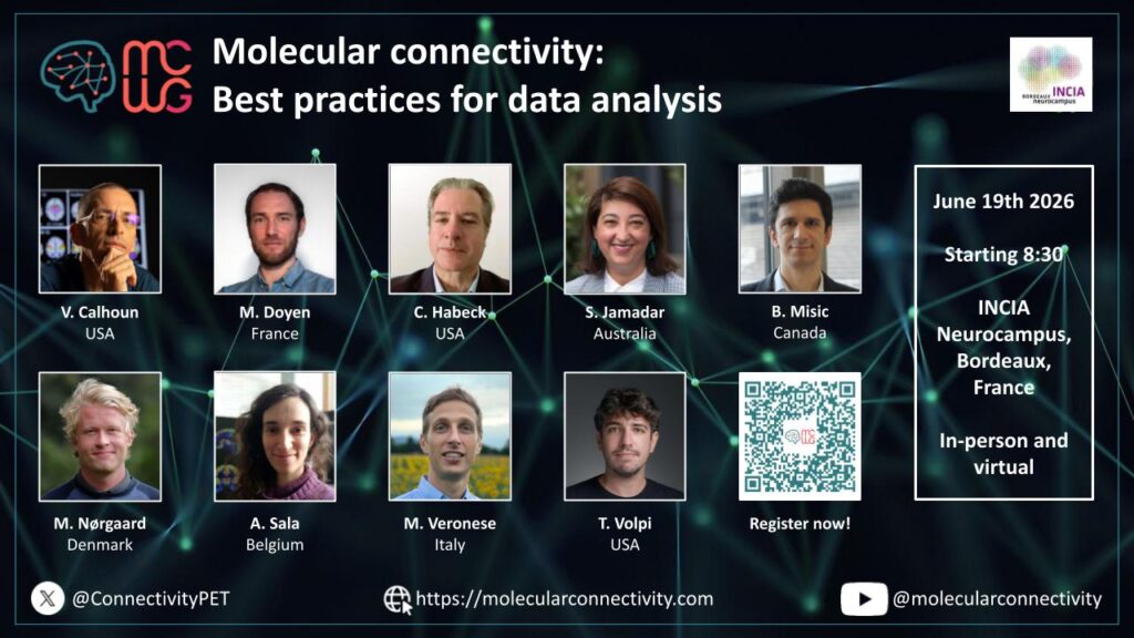
Molecular connectivity: Best practices for data analysis
Bordeaux June 19th, 2026 – (In-person or virtually)
Registration: Register Here! Free of Charge!
Program & Speakers:
8:30 – 08:40 Welcome & Introduction by the organizers
08:40 – 09:10 (30 min) Molecular connectivity in the broader context of fMRI and other modalities –Bratislav Misic, McGill University (Canada)
09:10 – 9:30 (20 min) Introduction to molecular connectivity and nomenclature in the context of the Delphi study – Sharna Jamadar, Monash University (Australia)
9:30 – 9:50 (20 min) Overview of commonly used methods for assessment of molecular connectivity with emphasis on technical aspects that require discussion – Mattia Veronese, University of Padua (Italy)
09:50 – 10:10 (20 min) Preprocessing: Data harmonization, PVC, normalization – Martin Norgaard, University of Copenhagen (Denmark)
10:10 – 10:30 Coffee break
10:30 – 10:50 (20 min) General prerequisites (population heterogeneity, statistical power), minimum number of subjects for inter- and intra-subject estimation – Arianna Sala, Université de Liège (Belgium)
10:50 – 11:10 (20 min) ROI-level estimation metrics: partial or Pearson correlation, Euclidean Similarity – Tommaso Volpi, Yale University (USA)
11:10 – 11:30 (20 min) Voxel-level estimation: SSM-PCA vs. ICA (which method and when? selection of components), seed-based correlation – Matthieu Doyen, Université de Lorraine (France)
11:30 – 11:50 (20 min) Best practices for merging molecular, functional information and clinical info – Vince Calhoun, GSU, GATech, Emory University (USA)
11:50 – 12:10 (20 min) Statistical robustness (bootstrapping, corrections) – Chris Habeck, Columbia University (USA)
12:10 – 13:00 Summary and plan for future steps
📝Network-based disease fingerprinting with neuroinflammation PET imaging
The goal of this study by Barzon and colleagues was to develop a data-driven, network-based approach to generate individual brain-wide TSPO PET matrices, employing Euclidean distance to quantify inter-regional pharmacokinetics similarity. They applied this approach to a large multicenter dataset of 528 PET scans utilizing three different TSPO tracers, including healthy controls and patients with different diseases.
Read the full study in Journal of Neuroinflammation.
Key Findings:
📝Personalized mapping of body homeostasis using whole-body PET connectomics and routine FDG PET imaging
Labarthe and colleagues introduced a framework for generating individualized PET-based connectomes, enabling robust assessment of personalized systemic homeostasis.
Read the full study in Communications Medicine.
Key Findings:
📝Metabolic brain connectivity analysis of a depressive-like phenotype in rats: a graph theory PET study
In this study, Vazquez-Matias and colleagues aimed to investigate whether there are metabolic connectivity alterations in the brain of rats with a depressive-like phenotype, using positron emission tomography (PET) and graph theory methods. Male Wistar rats were exposed to 5 days of repeated social defeat (RSD) to induce a depressive-like phenotype, and brain connectivity was assessed with [18F]FDG-PET.
Read the full study in Psychiatry Res.
Key Findings:
📝Neuro–visceral immunometabolic network phenotyping via baseline whole-body [¹⁸F]FDG PET/CT underlying pathologic response to neoadjuvant immunochemotherapy in resectable NSCLC
In this study by Shi and colleagues, whole-body FDG-PET was used to compare cerebral metabolic networks between non-small cell lung cancer patients with pathological response to immunotherapy and patients that did not respond, as well as the characterisation of the brain-visceral metabolic coupling. Brain regions were grouped into two pre-defined cerebral modules: the Warburg-related module (regions with consistently high physiological glucose metabolism), and the anxiety/stress-related module.
Read the full study in European Journal of Nuclear Medicine and Molecular Imaging.
Key Findings:
📝[18F]Fluorodeoxyglucose positron emission tomography ([18F]FDG PET) characterises neurodegeneration levels across the -synucleinopathy continuum
Orso and colleagues aimed at assessing the ability of previously described brain metabolic disease-related covariance patterns (RP) to characterise neurodegeneration across the alpha-synuclein continuum, as well as in the specific clinical phenotypes (i.e., cognitive- and motor-predominant). To this end, they included healthy controls and isolated rapid eye movement sleep behaviour disorder, Parkinson’s disease (PD), and dementia with Lewy bodies patients.
Read the full study in Movement Disorders.
Key Findings:
📝Mapping individual molecular connectomes in Alzheimer’s disease
In this study, Xu and colleagues characterised the individual variations in amyloid tau pathology across the Alzheimer’s disease continuum by building individual molecular connectomes that capture the unique variations in pathological deposits in every patient. This new proposed measure of connectome alteration shows high discriminative ability both at baseline and in longitudinal analyses and correlates with cognitive decline over time more strongly than conventional SUVR metrics.
Read the full study in Alzheimer’s and Dementia.
Key Findings:
The MCWG Outreach Council invites you to submit announcements or information about papers, conferences, presentations or other events or news related to brain and molecular connectivity as well as any positions available or job opportunities that you wish to publicize and share with the community!
Please submit any material for consideration by the final day of each month using this form – thank you!

The MCWG is made up of four international and multidisciplinary councils dedicated to promoting molecular connectivity research via dissemination of methods, results, collaboration, and resource sharing (e.g. datasets, tools) within the scientific community. We encourage the neuroscientific community to take an integrative perspective in study of the brain connectome, where various methods including MRI-based techniques, electrophysiological tools, and molecular imaging advance our understanding of the brain. Please find fundamental questions outlined here: “Brain connectomics: time for a molecular imaging perspective?”
Our website can be found here. We also invite you to join the MCWG!


