
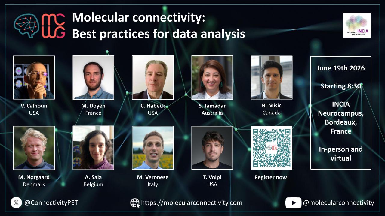
Molecular Connectivity: Best practices for data analysis
Bordeaux, June 19, 2026
We are excited to announce our second international symposium on Molecular Connectivity, entitled “Molecular connectivity: Best practices for data analysis”.
The symposium will be held on June 19th 2026 at Bordeaux (France), at Neurocampus facilities immediately following the Organization for Human Brain Mapping (OHBM) annual meeting. It builds on the success of our previous events: a satellite meeting held during the Brain & Brain PET conference in Glasgow in May 2022 (374 participants – 50 in person and >1600 views on Youtube), and a first dedicated symposium held in Munich in May 2024 (242 participants – 60 in person). Both events were very successful, led to the formation of our Molecular Connectivity Working Group which holds regular online seminars, and has produced influential papers [1][2].
We have assembled a superb list of speakers with the preliminary program listed below. The symposium will provide a forum for a vibrant exchange of ideas with the aim of significantly contributing to defining best practices in data analysis in this rapidly evolving field. Please join us in this very important effort. Any updates will be posted on our website and communicated in the next newsletter – stay tuned!
The MCWG organizes free monthly online seminars on brain connectivity and molecular imaging (see below).
Our online serie’s aim is to include the latest research findings from recently published papers on molecular connectivity. We will offer tutorials on methods and research resources for molecular connectivity estimation and we will discuss relevant findings in the field of brain connectivity that could aid study design and methodological development in the field of molecular connectivity.
The seminar will comprise a 30 minute presentation followed by discussion (~25 minutes).
Jan’24 MCOS001: Brain connectomics: Time for a molecular imaging perspective? A. Sala, Liège, Belgium.
Date: January 19th, 2024, Time: 15:00 CET, 9:00 EST
Please register here.
Abstract: In the past two decades brain connectomics has evolved into a major concept in neuroscience. However, the current perspective on brain connectivity and how it underpins brain function relies mainly on the hemodynamic signal of functional magnetic resonance imaging (MRI). Molecular imaging provides unique information inaccessible to MRI-based and electrophysiological techniques. Positron emission tomography (PET) has been successfully applied to measure neural activity, neurotransmission, and proteinopathies in normal and pathological cognition. In this talk, I position molecular imaging within the brain connectivity framework from the perspective of timeliness, validity, reproducibility, and resolution. I argue that the neuroscientific community should take an integrative approach whereby MRI-based and electrophysiological techniques, and molecular imaging contribute to our understanding of the brain connectome.
Biosketch: Arianna Sala is currently a FNRS post-doctoral research fellow at the Coma Science Group, University of Liège, Belgium. After a PhD in molecular medicine investigating molecular biomarkers in Alzheimer’s disease, she joined the Coma Science Group to study brain glucose metabolism and metabolic networks in disorders of consciousness. She is co-founder of the Molecular Connectivity Working Group, a multidisciplinary initiative that aims to evaluate molecular imaging for the study of brain connectivity.
Feb’24 MCOS002: Basic introduction to multivariate neuroimaging analysis – for nerds and novices C. Habeck, New York, United States.
Date: February 23rd, 2024, Time: 15:00 CET, 9:00 EST
Please register here.
Abstract: I will (1) go through some of the basics of PCA with toy simulations, (2) discuss some of the recent criticisms of PCA, and (3) show examples in fMRI data to demonstrate the power of multivariate compared to univariate analysis. I hope to convince the audience that an intelligent use of PCA is the best go-to initial pass at complex data, and also provides a good benchmark for more complicated and deeper learning architectures.

Biosketch: Chris Habeck was originally trained as a Particle Physicist, but followed the great migration from Physics to the lifesciences after his PhD. After a brief stint of biophysical computational modeling at the Neurosciences Institute in San Diego, he came to brain imaging analytics with PET and fMRI at Columbia University where he has been ever since. Chris has conducted, developed, and popularized multivariate analysis frameworks and non-parametric statistics, relying on the trusty workhorse of PCA: “My hope is that every practitioner -from seasoned analyst to novice- will get something out of my talk.“
Mar’24 MCOS003: NeuroMark PET: Towards a fully automated PET ICA pipeline V. Calhoun, Atlanta, United States.
Date: March 22nd, 2024, Time: 15:00 CET, 10:00 EST
Abstract: Anatomy based atlases are often used to summarize positron emission tomography (PET) data. However, it is well known that functional boundaries do not correspond well to anatomic boundaries. In addition, anatomic atlases do not capture variation among individuals and may average together voxels which are functionally distinct. In contrast, independent component analysis (ICA) provides informative data-driven output which also allows for overlap across anatomic regions. However, the output of blind ICA, without any a priori information, can be challenging to compare across studies and requires manual selection of components of interest. Here we propose the use of a spatially constrained ICA approach, called NeuroMark PET, leveraging a priori PET template derived from ICAs which replicated across independent PET datasets. We use this to generate a radioligand specific template which is then used to automatically generate component maps (i.e., covarying networks) and subject specific output using spatially constrained ICA. We also show that the ICA component maps are invariant to standard update value ratio (SUVR) scaling, allowing easy rescaling of the component loadings as desired. The proposed Neuromark PET approach effectively captures biologically meaningful subject-specific features that are comparable across different individuals. We demonstrate by comparing group differences in PET networks as well as age-related changes. This approach also allows for comparison across modalities or radioligands. We show an example of this by using a widely used functional MRI NeuroMark template as spatial priors for PET data, revealing PET specific modularity in fMRI intrinsic networks. In sum, we describe a PET NeuroMark template and pipeline and suggest that it represents a powerful resource for the research community and for analyses with large multimodal datasets.
Biosketch: Dr. Calhoun is founding director of the tri-institutional Center for Translational Research in Neuroimaging and Data Science (TReNDS) where he holds appointments at Georgia State, Georgia Tech and Emory. He is the author of more than 1000 full journal articles. His work includes the development of flexible methods to analyze neuroimaging data including blind source separation, deep learning, multimodal fusion and genomics, neuroinformatics tools. Dr. Calhoun is a fellow of the Institute of Electrical and Electronic Engineers, The American Association for the Advancement of Science, The American Institute of Biomedical and Medical Engineers, The American College of Neuropsychopharmacology, The Organization for Human Brain Mapping (OHBM) and the International Society of Magnetic Resonance in Medicine. He currently serves on the IEEE BISP Technical Committee and is also a member of IEEE Data Science Initiative Steering Committee as well as the IEEE Brain Technical Committee.
Apr’24 MCOS004: The many faces of brain connectivity S. Eickhoff, Jülich, Germany. Cancelled
Date: April 19th, 2024 Time: 15:00 CET, 9:00 EST
Regional segregation and long-range integration are the two fundamental principles of brain organization. As connectivity patterns play a major role in both of these, assessing the the connectome has emerged as a key avenue towards a better understanding of the brain. Key to this presentation is the notion that there is no such thing as “the” connectivity between two brain regions, but rather different concepts, facets and assessment of brain connectivity may provide corroborating, complementary or conflicting evidence. Therefore, a better understanding of the relationship between connectivity-types and their integration to a more holistic view of long-range interactions should be critical to avoid an undue reliance on individual approaches that may actually hinder progress in neurobiological understanding.
Simon Eickhoff is a full professor and chair of the Institute for Systems Neuroscience at the Heinrich-Heine University in Düsseldorf and the director of the Institute of Neuroscience and Medicine (INM-7, Brain and Behavior) at the Forschungszentrum Jülich. He is furthermore a visiting professor at the Chinese Academy of Science Institute of Automation. Working at the interface between neuroanatomy, data-science and brain medicine, he aims to obtain a more detailed characterization of the organization of the human brain and its inter-individual variability in order to better understand its changes in advanced age as well as neurological and psychiatric disorders. This goal is pursued by the development and application of novel analysis tools and approaches for large-scale, multi-modal analysis of brain structure, function and connectivity as well as by machine-learning for single subject prediction of cognitive and socio-affective traits and ultimately precision medicine.
May’24 MCOS005: Individual PET connectomes capture disease progression and cognitive decline in Alzheimer’s disease J. Pereira, Stockholm, Sweden.
Date: May 17th, 2024 Time: 15:00 CET, 9:00 EST
Identifying unique differences is crucial for enhancing personalized medicine strategies for Alzheimer’s disease (AD). In this work, we present a framework to create individual molecular connectomes by using longitudinal data from tau and amyloid positron emission tomography scans. We show that these personalized molecular connectomes can pinpoint specific individuals and track disease progression throughout different AD stages as well as over time.

I have a background in neuroimaging in aging and neurodegenerative diseases, mainly Alzheimer’s disease (AD). In my group we are working with different brain imaging modalities, CSF and plasma biomarkers to understand the sequence of pathological events that lead to AD. We have developed different methods, including an open-access software for the analysis of brain connectivity using graph theory, multilayer network approaches and deep learning called BRAPH.
Jun’24 MCOS006: Molecular connectivity & dynamic PET: comparing time series and subject series approaches T. Volpi, New Haven, Connecticut, United States.
Date: June 14th, 2024 Time: 15:00 CET, 9:00 EST
Please register here.
Dynamic PET acquisitions allow individuals to obtain individual estimates of molecular connectivity (MC) from time series data – similarly to fMRI functional connectivity. In this talk, focusing on [18F]FDG as a representative tracer, I will discuss
Much of this content is included in our 2023 JCBFM paper.

Dr. Tommaso Volpi is currently a Postdoctoral Associate at the Department of Radiology at Yale University, under the supervision of Prof. Richard Carson. He obtained his Ph.D. in 2023 from the University of Padova, Italy, with a thesis on the complex coupling between [18F]FDG PET measures of brain glucose metabolism and fMRI proxies of spontaneous activity, including the comparison of metabolic and functional connectivity. His main current projects concern methods for PET kinetic modeling and noninvasive input function estimation to obtain physiologically informative parameters, and the integration of PET- and MRI-derived features to understand brain function and connectivity in health and disease (Parkinson’s disease, epilepsy, gliomas). He is a member of the Validation Council of the MCWG.
You can find more about Tommaso here!
Oct’24 MCOS007: Test-retest reproducibility of structural and proxy estimates of brain connectivity at rest I. Yakushev, Munich, Germany.
Date: October 18th, 2024 Time: 15:00 CET, 9:00 EST
Please register here.
While structural connectivity (SC) is indicative of actual anatomical connectivity and derived at a single subject level, proxy estimates of brain connectivity such as functional connectivity (FC) from functional magnetic resonance imaging (MRI), intersubject covariance of regional gray matter volume (GMVcov) from structural MRI, and intersubject covariance of regional 18F-fluorodeoxyglucose uptake (FDGcov) from positron emission tomography are derived from statistical dependencies between regional measurements. To understand, in how far these estimates are able to capture physiological and pathological changes in brain connectivity, knowledge on their reproducibility is essential. In this study, we determined reproducibility of group level SC, FC, GMVcov, and FDGcov in the same 55 healthy subjects at rest using a simultaneous PET/MRI acquisition protocol. Reproducibility was determined using Spearman’s correlation coefficient, coefficient of variation, and proportion of repeatedly (test and retest) present connections.

Igor Yakushev obtained his MD in 2010 from the University of Mainz, Germany. In 2012, after residency in Psychiatry and Neurology, he moved to Munich for residency in Nuclear Medicine at the Technical University of Munich (TUM), Germany. Since 2013 he has led the research group “Multimodal imaging of normal and pathological cognition”. Since 2015 he is the Head of Neuroimaging, since 2018 Senior Consultant in Nuclear Medicine at the Dept. of Nuclear Medicine, TUM. He is an Associate Faculty at the Munich Center for NeuroSciences “Brain and Mind”. Igor Yakushev has served in the Board of Directors of the Brain Imaging Council, Society of Nuclear Medicine and Molecular Imaging, as well as in the Neuroimaging Committee, European Association of Nuclear Medicine. His research is focused on mechanisms of brain connectivity and development of imaging-based biomarkers for neurodegenerative and neuro-oncological disorders.
You can find more about Igor here!
Nov’24 MCOS008: Metabolic connectivity in ageing H. Deery, Melbourne, Australia.
Date: November 22nd, 2024 Time: 14:00 CET, 8:00 EST, 22:00 AEST
Please register here.
Information transfer across the brain has a high energetic cost and requires efficient glucose metabolism. Here we use recently developed high temporal resolution functional positron emission tomography (fPET) to create a timecourse of glucose metabolism for individual subjects and assess the relationship between metabolic connectivity and cognitive function in ageing. The metabolic connectomes of 40 younger (mean 27.9 years; range 20-42) and 46 older (mean 75.8; 60-89) adults were characterised by high connectivity in the frontal, temporal, motor, parietal and medial cortices. Older age was associated with lower global integration of metabolic hub regions. In younger adults, a high proportion of glucose was used to support hubs in the frontal regions. Older adults used a higher proportion of a smaller energy budget to support mostly posterior hub regions. This metabolic network topology of older adults was associated with worse cognitive performance. We conclude that ageing is associated with a high glucose cost in hub regions and disrupted information transfer across the metabolic network. Our results highlight the fundamental role that metabolism plays in supporting information transfer in the brain and the unique insights that metabolic connectivity provides into the ageing brain.

Hamish Deery is a Ph.D. candidate in the School of Psychological Sciences and the Cognitive Neuroimaging Lab led by A/Professor Sharna Jamadar at Monash Biomedical Imaging, Monash University, in Melbourne, Australia. His research interests are in understanding brain function and cognition across the adult lifespan, including unique and potential shared pathways in ageing and lifestyle diseases. His work includes the use of simultaneous functional PET and MR to investigate the impact of insulin resistance on cerebral gluose metabolism and cognition in mid- and later-life.
Jan’25 MCOS009: Epigenetic alterations in white matter with age: impacts on structural connectivity and beyond M. Catanese, Boston, Massachusetts, United States.
Date: January 17th, 2025 Time: 15:00 CET, 9:00 EST
Please register here.
Age related structural change in the healthy human brain has been associated with cognitive decline, however associated molecular alterations remain poorly understood. Positron emission tomography (PET) enables the in vivo investigation of molecular targets and can accelerate therapeutic development through their quantification. It has recently become possible to measure epigenetic machinery in the living human brain using the PET radiotracer [11C]Martinostat. This imaging agent targets a subset of class I histone deacetylases or HDACs. Epigenetic deacetylation of lysine residues of histone tails modifies chromatin in the regulation of gene expression. I am interested in HDACs as they are heritable and play critical roles in our response to the environment and are important in relation to cognitive processes including learning, memory and synaptic plasticity. Moreover, HDACs are implicated in psychiatric and neurodegenerative disorders. In previous work an age-related increase in [11C]Martinostat uptake was observed with age in healthy individuals and localized to the white matter. This increase in uptake correlated and co-localized with decreased structural integrity. I will discuss my goal to extend this work to study epigenetic alterations in relation to major depressive disorder by examining the potential association of epigenetic alterations (altered uptake) in relation to symptom related networks. I will also discuss using imaging-guided postmortem research programs initiated for the dual purpose of informing data interpretation and interrogating HDAC-related biology. My ultimate goal is to help better understand molecular dysregulation in psychiatric disorders.

Mary Catanese is a Faculty Instructor in the Department of Psychiatry Department of Psychiatry, Massachusetts General Hospital, Harvard Medical School. She received her Ph.D. from the University of Massachusetts Amherst. Her work is focused on better understanding molecular dysregulation in psychiatric disorders using a multidimensional approach, integrating neuroscience, neuroimaging and clinical questions to address patient needs.
Feb’25 MCOS010: Mapping brain function and connectivity in rodents using small animal PET/MRI K. Herfert, University Hospital of Tuebingen, Germany.
Date: February 21st, 2025 Time: 15:00 CET, 9:00 EST
Please register here.
Simultaneous small-animal PET/MRI integrates molecular and hemodynamic measures to offer deeper insights into brain function and connectivity. In our recent study, we combined functional PET (fPET) and fMRI to investigate the nigrostriatal pathway in rats under optogenetic stimulation, revealing pronounced inhibition in specific downstream regions rather than widespread activation. This indicates an unexpected inhibitory regulation within this key motor circuit. In another study, we compared molecular and functional connectivity measures following MDMA administration in rats. By pairing PET-based imaging of molecular targets with BOLD fMRI, we uncovered complementary insights into how neurotransmitter systems influence large-scale networks. These results underscore the power of fPET/fMRI to detect subtle changes in neuronal and molecular processes, highlighting the added value of multimodal imaging in shaping our understanding of brain function and informing both basic neuroscience and clinical research.

Prof. Dr. rer. nat. Kristina Herfert is Associate Professor for Functional and Metabolic Brain Imaging at the Werner Siemens Imaging Center, University of Tübingen, Germany. A trained neuroscientist, she specializes in in vivo PET quantification of dopaminergic and serotonergic receptors, PET tracer development for brain protein aggregates, and simultaneous small-animal PET/fMRI to study molecular and functional networks. After completing her doctorate under Prof. B.J. Pichler, she was awarded a Research Fellowship in Prof. Feng Zhang’s lab at the Broad Institute of MIT and Harvard. Professor Herfert’s research uniquely bridges gene editing approaches with neuroimaging to understand the brain’s molecular, cellular, and network-level functions.
Please find further information about Dr. Herfert here!
Mar’25 MCOS011: Comparing Intra- and Inter-individual Correlational Brain Connectivity from Functional and Structural Neuroimaging Data X. Di, New Jersey Institute of Technology, United States.
Date: March 21st, 2025 Time: 15:00 CET, 10:00 EDT
Please register here.
Many neuroimaging modalities, such as PET and structural MRI, typically assess connectivity using inter-individual correlations, although these measures may be affected by several factors. To clarify these influences, we compared intra- and inter-individual correlations using identical data types. Our analysis of two distinct datasets revealed that long-term age effects contribute to correlations in brain structure, though they bear limited resemblance to functional connectivity. In contrast, both intra- and inter-individual correlations in functional measures (PET and ReHo) strongly reflected functional connectivity, underscoring the importance of state-like brain activity. These findings highlight the need to account for such influences when interpreting inter-individual connectivity measures.
Dr. Xin Di is a Research Assistant Professor in the Department of Biomedical Engineering at the New Jersey Institute of Technology. With a PhD in psychology, his research focuses on modeling brain connectivity using multi-modal neuroimaging data, including fMRI, PET, structural MRI, and fNIRS. He also investigates brain alterations associated with autism spectrum disorder.
Please find further information about Dr. Di here!
Apr’25 MCOS012: Molecular Connectivity in Neurotransmission M. Veronese & M. Severino, University of Padua, Italy.
Date: April 11th, 2025 Time: 15:00 CET, 09:00 EDT
Please register here.
PET and SPECT are fundamental tools for studying neurotransmission in living systems. Traditionally, these imaging modalities are analysed by focusing on individual regions or voxels independently. However, recent interest has shifted toward molecular connectivity approaches, which offer a fresh perspective on the brain’s complex organization by examining interactions between brain regions. This talk will provide a comprehensive review of molecular connectivity studies in neurotransmission, highlighting their expanding applications, advantages over traditional methods, and contributions to advancing neuroscience.

Dr. Mattia Veronese is Associate Professor in Biomedical Engineering at the Department of Information Engineering, University of Padua and Honorary Senior Lecturer in Neuroimaging at King’s College London. He is a biomedical engineer by training and holds a PhD in PET kinetic modelling. His main research interest is related to the development and validation of molecular neuroimaging biomarkers and to their use for drug development and precision medicine.
Click here for more information about him.

Mario Severino is a PhD student in Bioengineering at the Department of Information Engineering, University of Padua. He holds a Master’s degree in Bioengineering. His primary research focus is on applying PET molecular connectivity and network science approaches, aiming to establish these methods as precision medicine biomarkers.
May’25 MCOS013: Great expectations but a bumpy road: Caveats in neuroimaging analyses and modelling S. Eickhoff, Heinrich-Heine University, Germany.
Date: May 23rd, 2025 Time: 15:00 CET, 09:00 EDT
Please register here.
The increasing availability of large cohorts and tools for multivariate statistical learning, allowing the prediction of individual cognitive or clinical phenotypes in new subjects, started a revolution towards neuroimaging applications in precision medicine. While the field has enjoyed a lot of enthusiasm recently, the road towards translation and re-life applications may be considerably more challenging than often acknowledged. Following a short overview on the motivation and perspectives, the major part of my talk will focus on several critical yet often underappreciated challenges for such endeavors. These include on the one hand technical and biological aspects that may undermine the validity of prediction results, due to the inherent low-dimensional structure of biological variability. On the other hand, ethical, legal and societal aspects will ultimately shape practical adaptation but need stronger consideration in the development of new pipelines if these are to move beyond proof-of-concept work.
Simon Eickhoff is a full professor and chair of the Institute for Systems Neuroscience at the Heinrich-Heine University in Düsseldorf and the director of the Institute of Neuroscience and Medicine (INM-7, Brain and Behavior) at the Forschungszentrum Jülich. He is furthermore a visiting professor at the Chinese Academy of Science Institute of Automation. Working at the interface between neuroanatomy, data-science and brain medicine, he aims to obtain a more detailed characterization of the organization of the human brain and its inter-individual variability in order to better understand its changes in advanced age as well as neurological and psychiatric disorders. This goal is pursued by the development and application of novel analysis tools and approaches for large-scale, multi-modal analysis of brain structure, function and connectivity as well as by machine-learning for single subject prediction of cognitive and socio-affective traits and ultimately precision medicine.
Sep’25 MCOS014: Simultaneous EEG-PET-MRI identifies temporally coupled, spatially structured hemodynamic and metabolic dynamics across wakefulness and NREM sleep. J. Chen, Martinos Center for Biomedical Imaging, United States.
Date: September 19th, 2025 Time: 15:00 CEST, 09:00 EDT
Please register here.
Sleep induces profound changes in cerebral hemodynamics and metabolism, yet how these processes unfold across wakefulness and sleep, as well as their spatiotemporal dependence remains largely unknown. In this talk, Dr. Chen will present her recent work integrating functional PET (fPET)-FDG with simultaneous EEG-fMRI to track physiological dynamics across the sleep–wake cycle. Findings show that global hemodynamic and metabolic signals are tightly coupled during the descent into NREM sleep, both closely tracking EEG arousal dynamics. There were identified two distinct network patterns in NREM sleep. These findings shed light on how sleep can diminish awareness while preserving sensory responsiveness, revealing a complex, alternating balance of hemodynamic and metabolic processes in the sleeping brain. This work also highlights the potential of EEG–PET–MRI for probing the neuro-hemo-metabolic underpinnings of cognition and arousal in humans.

Dr. Chen is an Assistant Professor at the Martinos Center for Biomedical Imaging at Massachusetts General Hospital and Harvard Medical School. She received her Ph.D. from Stanford University, with a major in Electrical Engineering and a minor in Statistics. Following graduation, she pursued further training in neuroimaging as a postdoctoral fellow at the Martinos Center. Her lab advances multimodal dynamic functional imaging techniques and computational approaches to investigate the biophysical and molecular mechanisms underlying intrinsic brain activity, as well as the neurobiological consequences of cognition, arousal, and disease.
Oct’25 MCOS015: Metabolic connectivity features in Alzheimer’s disease. S. Caminiti, University of Pavia, Italy.
Date: October 17th, 2025 Time: 15:00 CEST, 09:00 EDT
Please register here.
Abstract: Emerging evidence highlights that connectivity alterations in Alzheimer’s disease (AD) include not only hypo-connectivity, but also hyper-connectivity, particularly in limbic and cerebellar regions during the prodromal and early dementia stages. In a recent longitudinal multimodal study, it was demonstrated that hyper-connectivity is detectable with FDG-PET metabolic connectivity and is not merely compensatory. These findings converge with other recent reports showing that medial temporal and anterior-temporal hyper-connectivity mediates amyloid-related tau accumulation and accelerates neurodegeneration. Altogether, converging data suggest that hyper-connectivity, far from being a benign or compensatory feature, may actively drive AD progression by increasing metabolic burden and synaptic inefficiency. Understanding this dual landscape of hypo- and hyper-connectivity offers new insights into AD pathophysiology and may open perspectives for network-based biomarkers of disease progression.

Dr. Caminiti is an Assistant Professor at the Department of Brain and Behavioral Sciences at the University of Pavia in Italy. With a background in neuroimaging and neuroscience, she is dedicated to advancing our understanding of the intricacies of the brain and its functioning. Her primary research focus revolves around aging and neurodegenerative diseases. The goal of her research program is to examine selective neuronal vulnerability in Alzheimer’s and Parkinson’s disease using multimodal neuroimaging including PET and MRI.
Nov’25 MCOS016: Validating brain connectivity measures: integrating biological, statistical, and clinical evidence. A. Jasanoff (MIT, USA), A. Hildebrandt (University of Oldenburg, Germany), Z.-Q. Liu (McGill University, Canada), M. Perovnik (University of Ljubljana, Slovenia)
Date: November 21st, 2025 Time: 15:00 CET, 09:00 EST
Please register here.


Abstract: In this symposium, we will show how validation frameworks across biological, statistical and clinical dimensions help ground our interpretation of connectivity measures, establishing more reliable tools for both basic neuroscience and clinical applications. After a short introduction on the concept of validation by the organizers, Alan Jasanoff will tackle biological validation, showing how a novel genetically encoded activity reporter in animal models can inform the biological basis of fMRI brain connectivity. Second, Andrea Hildebrandt will show how a systematic characterization of the brain connectivity multiverse can provide valuable insights into assessment of statistical robustness across analytical choices as a prerequisite for enhancing replicability. Third, Zhen-Qi Liu will provide an overview of the properties of MR, MEG and PET brain connectivity measures, highlighting which metrics possess desirable statistical and biological properties. Last, Matej Perovnik will show a real-life example of systematic clinical validation based on PET brain networks.
Jan’26 MCOS017: Connectivity-based parcellation to map brain organization. S. Genon, University of Dusseldorf, Germany
Date: January 23rd, 2026 Time: 14:00 UTC
Please register here.
Abstract: The human brain is often described in terms of discrete regions, yet defining brain atlases remains a central challenge in neuroscience. Connectivity-based parcellation offers a principled framework for identifying functionally coherent regions using a variety of connectivity markers. In this talk, Dr. Sarah Genon will highlight how metabolic connectivity can be leveraged to derive region definitions grounded in metabolic network organization. I will discuss the relevance of these connectivity-based regions for improving our understanding of brain–behavior relationships and characterizing dysfunction in clinical populations.

Dr. Sarah Genon is a cognitive neuroscientist specialized in neuroimaging, machine learning, and the study of brain–behavior relationships. She is a Heisenberg Professor at the Heinrich-Heine University of Dusseldorf and a group leader at the Forschungszentrum Jülich (Germany).
Feb’26 MCOS018: Constructing the human brain metabolic connectome with MR spectroscopic imaging reveals cerebral biochemical organization. P. Klauser, F. Lucchetti, Lausanne University Hospital, Switzerland
Date: February 20th, 2026 Time: 14:00 UTC
Please register here.
Abstract: Fast three-dimensional proton magnetic resonance spectroscopic imaging (¹H-MRSI) now enables whole-brain mapping of metabolites, offering a biochemical dimension to brain organization that is largely missing from current connectomics. Using high-resolution ¹H-MRSI in 51 healthy participants, we construct a within-subject metabolic connectome defined by correlations among five metabolites (tCr, tNAA, Glx, Ins, Cho) across gray-matter regions. The resulting networks reveal a dominant metabolic similarity mode forming a continuous caudal-to-rostral gradient and which reflects a trade-off between local metabolic homogeneity. Metabolic similarity overlaps partly with structural hubs but aligns more strongly with cytoarchitectonic similarity and gene co-expression than with tractography-based connectivity. These findings establish metabolic similarity as a robust, biologically grounded axis of brain organization and position ¹H-MRSI as a complementary modality for connectomics in health and disease.

Paul Klauser, MD-PhD, is a child psychiatrist, consultant in the Department of Psychiatry at Lausanne University Hospital, tenure-track Assistant Professor at the University of Lausanne and Head of the Research Unit on Psychosis in Lausanne Switzerland. ORCID; LinkedIn.

Federico Lucchetti, PhD, is a trained physicist who earned his PhD in Neuroscience from the Free University of Brussels in 2019. He then spent several years at the University of Luxembourg, where he worked on cyber resilience and distributed systems, before joining in 2023 Paul Klauser’s group, where he now focuses on whole-brain MR spectroscopic imaging (MRSI) and metabolic connectomics.
Mar’26 MCOS019: Human Cerebral Cortex Organization Characterized by Functional PET-FDG “Metabolic Connectivity”. P. Du, École polytechnique fédérale de Lausanne, Switzerland
Date: March 20th, 2026 Time: 14:00 UTC
Please register here.
Abstract: Resting-state metabolic connectivity (RSMC) as measured by [18F]-fluorodeoxyglucose functional PET (fPET-FDG) provides a promising framework for understanding the brain’s energetic organization, yet its spatiotemporal structure remains insufficiently characterized. Here, we investigated the cortical organizational principles of RSMC in the human brain and their relationship to other known cortical organizational features. At the local scale, metabolic boundaries partly overlapped with resting-state functional connectivity patterns. At the global scale, RSMC was organized along a robust superior-inferior cortical gradient, and was primarily driven by low-frequency, minute-scale fPET-FDG dynamics. This large-scale metabolic profile aligned with known anatomical and energetic constraints, providing new insights into the brain’s energetic architecture. Link to BioRxiv.

Penghui Du is currently a Master’s student in Neuro-X at EPFL. He received his BSc in Intelligent Medical Engineering from the Southern University of Science and Technology in 2024. As a visiting student at the CANDY Lab (Athinoula A. Martinos Center for Biomedical Imaging, Massachusetts General Brigham, United States), he worked with Dr. Jingyuan Chen on mapping the organization of the human cerebral cortex using functional PET-FDG metabolic connectivity. Personal website: https://penghui-du.com/
Apr’26 MCOS020: Preoperative network activity predicts the response to subthalamic DBS for Parkinson’s disease. P. Unadkat, Northwell Health, USA
Date: April 17th, 2026 Time: 13:00 UTC
Please register here.
Abstract: Does the brain’s baseline network architecture tell us about an individual’s capacity to respond to neuromodulation? Can we use this to identify which Parkinson’s disease patients will benefit most from deep brain stimulation before they ever reach the operating room? This talk will present our work developing and validating STN StimNet, a treatment-specific metabolic brain network identified in PD patients with implanted subthalamic nucleus (STN) electrodes. Several key questions will be addressed: What distinguishes a treatment-induced network from a disease-related one? What is the electrophysiological basis of this network; specifically, why do STN theta-band oscillations, rather than the pathological beta activity traditionally linked to PD motor impairment, drive StimNet expression? We go on to demonstrate how preoperatively measured network expression with either FDG PET or resting-state fMRI predicts postoperative motor outcomes, and how it compares to the current gold standards for patient selection. Using data from a large cohort of PD patients spanning the clinical spectrum of the disease, we illustrate how these measurements can stratify patients by their likelihood of achieving a clinically meaningful response, while appropriately excluding populations unlikely to benefit, such as those with atypical parkinsonian syndromes. Finally, this predictive framework may have relevance beyond STN-DBS in Parkinson’s disease. The principle that preoperative circuit-level activity shapes an individual’s response to focal neuromodulation could guide patient selection across other DBS targets and may extend to other movement and psychiatric disorders.

Dr. Prashin Unadkat, MBBS, PhD is the Chief Resident in the Department of Neurosurgery at the Zucker School of Medicine at Hofstra/Northwell and an incoming Stereotactic and Functional Neurosurgery fellow at Baylor College of Medicine. His clinical practice focuses on image-guided and neuromodulatory surgical strategies for movement disorders, psychiatric conditions, and epilepsy. His research centers on developing functional and structural brain imaging biomarkers that can predict treatment response and improve surgical planning, with a particular emphasis on network-level analysis of deep brain stimulation effects.
May’26 MCOS021: Deciphering the biological correlate of metabolic connectivity in neurodegenerative diseases. J. Gnörich, LMU Munich, Germany
Date: May 22nd, 2026 Time: 13:00 UTC
Please register here.
Abstract: Metabolic connectivity derived from [18F]FDG PET is an emerging network-based biomarker in neurodegenerative diseases, yet its underlying biological basis remains insufficiently understood. In this talk, I will present our recent published data combining [18F]FDG-PET with scRadiotracing, demonstrating that metabolic connectivity is strongly influenced by cell-specific glucose uptake patterns of neurons, astrocytes, and microglia. Our findings identify astroglial metabolism as a key driver of age-related network reorganization and show that microglial presence and activation states substantially modulate cerebral metabolic networks in translational mouse models. In addition, I will present our ongoing research and preliminary data using single-cell transcriptomics to further dissect the molecular and subcellular mechanisms underlying metabolic connectivity. Together, these approaches aim to establish metabolic connectivity as a biologically informed imaging marker for neurodegenerative disease.

Dr. Johannes Gnörich is a clinician scientist at the Department of Nuclear Medicine, LMU University Hospital, LMU Munich, Germany. His research focuses on PET imaging, neuroinflammation, and biomarker development in neurodegenerative diseases. Since his doctoral thesis, he has published several translational studies, including methodological evaluations of single-cell radiotracer detection after in vivo tracer injection (scRadiotracing), aiming to disentangle the cellular sources of PET signals in different inflammatory disorders, including neurodegeneration. These studies contributed to the validation and establishment of novel PET tracers in the field. In addition, Dr. Gnörich integrates advanced network-based imaging analyses, particularly metabolic connectivity approaches, to characterize system-level alterations in brain diseases. Moreover, Dr. Gnörich has a strong record of coordinating academic research projects, including recent multi-center studies. He is actively engaged in international collaborations, serves as a peer reviewer for leading scientific journals, and mentors early-career researchers.
Please check out the following events during OHBM 2024:
9:00-13:00 CEST, May 3rd 2024
Free registration
TranslaTUM, Einsteinstraße 25, 81675 Munich, Germany and Streamed Live
The symposium is part of the event “Molecular Imaging of Brain Connectivity: towards standardized nomenclature“
May 28th, 2022
Glasgow, UK and Streamed Live
Free Registration
Free Live Streaming
Travel Bursaries Available to
Early-career Researchers
All talks of this event are available virtually here: Brain and Brain PET 2022 – Satellite Symposium
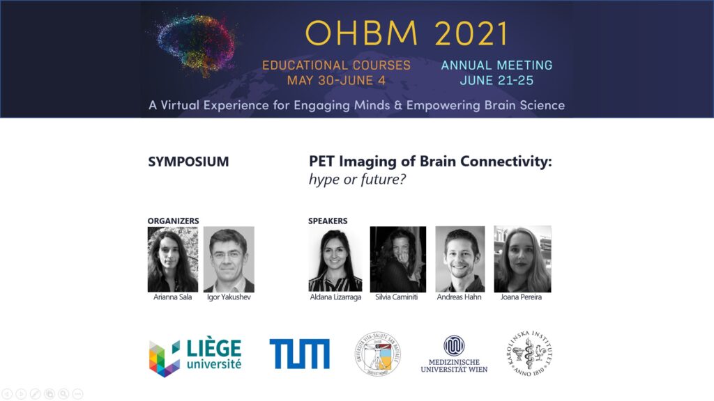
All materials of this OHBM 2021 symposium are available virtually here: OHBM 2021
OHBM membership, a previous registration to an OHBM conference or registration to the upcoming OHBM conference are required to access the materials.