
Thank you to everyone who joined us for April’s MCOS with Dr. Johannes Gnörich! Missed it? The recording is available here.
As the holiday season approaches, we would like to let our community know that there will be a short summer pause in our regular online MCOS activities. This break offers an opportunity to rest, reflect, and prepare for an exciting new season ahead.
We look forward to returning in September 2026 with a fresh series of online talks, discussions, and community conversations. Thank you for your continued support and participation throughout the year. We wish everyone a relaxing and enjoyable summer holiday season and look forward to reconnecting with you soon.
Each month, we will feature a member of the MCWG and have a brief Q&A!
This month please enjoy our highlight of Dr Tang, member of the MCWG Resource Council.

Dr Tang completed her undergraduate studies in Biomedical Engineering (Sun Yat-Sen University, China) and a master’s degree at Technical University of Munich, Dr Tang obtained her PhD in Medical Image Analysis at KU Leuven under the supervision of Prof. Dr. Michel Koole. Her research aims to advance quantitative brain PET imaging through the integration of machine learning and established PET kinetic modelling, non-invasive quantification, supervised classification, and network analysis.
Dr Tang has graciously responded to our feature questionnaire:
What sparked your interest in molecular imaging or led you to focus on research in molecular imaging?
My interest in molecular imaging began when I wrote my Master’s thesis, where I was exposed to various medical imaging modalities and their applications in healthcare. I think what initially attracted me to molecular imaging was the fact that it allows us to look beyond anatomy and actually observe biological processes in vivo. I found it remarkable that techniques such as PET can reveal pathology-specific changes at a molecular level, sometimes before structural abnormalities become apparent. As I learned more about the field, I became increasingly interested in the quantitative aspects of molecular imaging. I was drawn to the challenge of extracting meaningful biological information from complex imaging data and translating that information into clinically useful biomarkers. This interest eventually led me to focus on brain PET imaging and later to my work on machine learning, kinetic modelling, and quantitative analysis. I see great potential in combining advanced computational techniques with domain knowledge from molecular imaging to improve the accuracy, robustness, and clinical utility of imaging biomarkers. What continues to motivate me is the opportunity to contribute to both methodological innovation and, ultimately, better patient care.
What is your role in the Molecular Connectivity Working Group, and what have you been contributing to or working on within the group?
I’m a junior member of the Molecular Connectivity Working Group and part of the Resource Council. One of my main contributions has been helping to systematically review the latest research in molecular connectivity. We also collect information on the latest connectivity analysis methods and software tools so that researchers can more easily access and compare available approaches. My role allows me to stay up to date with new developments in molecular connectivity while contributing to community resources that support researchers working in this field
What do you think are the most important challenges in current brain connectivity research, or which unsolved/underappreciated issues should the community address?
I think one important challenge is the lack of signal-specific atlases for molecular connectivity analysis. We now have a growing number of PET tracers that target different biological processes, such as glucose metabolism, neurotransmitter systems, neuroinflammation, and proteinopathies. However, many studies still rely on brain atlases that were originally developed for structural or functional imaging. These anatomically defined parcellations may not accurately reflect the spatial organization of the molecular signals being measured. Developing tracer-specific or biologically informed atlases could potentially improve the sensitivity and interpretability of molecular connectivity analyses.
A second challenge is the interpretation of connectivity measures. In molecular connectivity studies, we often identify patterns of correlated tracer uptake across brain regions, but the biological meaning of these connections is not always clear. Unlike structural connectivity, which reflects physical white-matter pathways, or functional connectivity, which is linked to synchronized neural activity, molecular connectivity can arise from multiple mechanisms. For example, it may reflect shared receptor distributions, common metabolic demands, disease propagation pathways, or coordinated regulation of molecular processes. As a result, it is often difficult to determine what a connectivity pattern actually represents biologically.
Another underappreciated issue is validation. Many connectivity methods can produce interesting networks, but it is often difficult to establish a biological ground truth. Developing strategies to validate molecular connectivity measures across datasets, tracers, and disease populations will be important for ensuring that these findings are robust and clinically meaningful.
What scientist or scientific achievement do you most admire?
I would say Fei-Fei Li. Of course, I admire her scientific contributions to computer vision and AI, which have had a profound influence on medical image analysis. But what I admire even more is her view that AI should be developed with the right motivation. One quote from her that has stayed with me is: “The true impact of AI on the world would be largely determined by the motivation that guided the development of the technology.” As someone working at the intersection of AI and healthcare, I think that’s a very important reminder that our goal is not just to develop more advanced algorithms, but to create tools that ultimately address clinical needs.
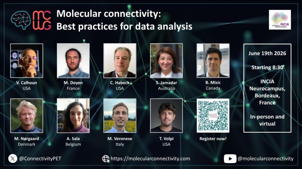
Molecular connectivity: Best practices for data analysis
Bordeaux June 19th, 2026 – (In-person or virtually)
Registration: Register Here! Free of Charge!
Registration closes on June 14th for in-person attendance and on June 17th for virtual attendance.
Program & Speakers (updated):
8:30 – 08:40 Welcome & Introduction by the organizers (Matthieu Doyen, Igor Yakushev)
Chairs: Joachim Mazere, Arianna Sala
08:40 – 09:10 (30 min) Molecular connectivity in the broader context of fMRI and other modalities
Bratislav Misic, McGill University (Canada)
09:10 – 9:30 (20 min) Introduction to molecular connectivity and nomenclature (Delphi study)
Sharna Jamadar, Monash University (Australia)
9:30 – 9:50 (20 min) Methods for estimation of molecular connectivity with emphasis on technical aspects
Mattia Veronese, University of Padua (Italy)
09:50 – 10:10 (20 min) Preprocessing: Data harmonization, PVC, normalization
Martin Nørgaard, University of Copenhagen (Denmark)
10:10 – 10:30 Coffee break
Chairs: Xin Di, Martin Norgaard
10:30 – 10:50 (20 min) General prerequisites; population heterogeneity, statistical power, minimum number of subjects for inter- and intra-subject estimation
Arianna Sala, Université de Liège (Belgium)
10:50 – 11:10 (20 min) ROI-level estimation metrics: partial or Pearson correlation, Euclidean Similarity
Tommaso Volpi, Yale University (USA)
11:10 – 11:30 (20 min) Voxel-level estimation: SSM-PCA vs. ICA: which method and when? Selection of components, seed-based correlation
Matthieu Doyen, Université de Lorraine (France)
11:30 – 11:50 (20 min) Fusion of functional, structural and clinical information
Vince Calhoun, GSU, GATech, Emory University (USA)
11:50 – 12:10 (20 min) Statistical robustness; bootstrapping, corrections
Chris Habeck, Columbia University (USA)
12:10 – 13:00 Summary (Igor Yakushev, Arianna Sala, Joachim Mazere, Mattia Veronese, Matthieu Doyen)
13:00 – 14:00 Network Lunch
Multimodal Integration in Human Brain Mapping
Bordeaux June 14, 2026 – (In-person at Palais 2 l’Atlantique – duration: 4h)
Organizers: Prof Dr Joana B Pereira and Dr Arianna Sala
Capturing Rich Multimodal Brain Interactions: Model Selection, Interpretability, and Clinical Applications (20 min)
Prof. Jing Sui – IDG/McGovern Institute for brain research, State Key Laboratory of Cognitive Neuroscience and Learning, Beijing Normal University, China
Neuromaps: A Python Toolbox for Cross-Modal Standardization and Interpretation (20 min)
Dr. Vincent Bazinet – Montréal Neurological Institute, McGill University, Montréal, Quebec, Canada
Linking MRI-Derived Measures and Underlying Neurophysiology Using the JuSpace Toolbox (20 min)
Prof. Juergen Dukart – Institute of Neurosciences and Medicine, Brain & Behaviour (INM-7), Research Centre Juelich; Juelich, Germany
Multilayer Network Modelling with the BRAPH 2 software (20 min)
Assoc. Prof. Joana B. Pereira – Clinical Neurosciences Department, Kalinska Institute, Stockholm, Sweden
Integrating fMRI and PET data through REACT (20 min)
Dr. Manuela Moretto – Department of Information Engineering, University of Padua, Padua, Italy
JuSpace: Spatial Correlation Analyses for Molecular–MRI Integration (35 min)
Prof. Juergen Dukart – Institute of Neurosciences and Medicine, Brain & Behaviour (INM-7), Research Centre Juelich; Juelich, Germany
Practical Tutorial with Neuromaps (35 min)
Dr. Vincent Bazinet – Montréal Neurological Institute, McGill University, Montréal, Quebec, Canada
Multilayer Network Analyses with BRAPH 2 (35 min)
MS Yu-Wei Chang – Department of Physics, University of Gothenburg, Gothenburg, Sweden
Receptor-Enriched Functional Connectivity with REACT (35 min)
Dr. Manuela Moretto – Department of Information Engineering, University of Padua, Padua, Italy
Event link: OHBM 2026
🎤 Fernando Bravo et al. – Insights from animal models: simultaneous fPET/fMRI during optogenetic and pharmacological interventions – Oral presentation.
Educational course “A glimpse into brain metabolic dynamics: An introduction to functional PET and multi-modal fusion”. Sunday 14th June from 9am to 1pm.
Event link: NRM 2026
🎤 Leonardo Barzon et al. – Characterising spatial disease signatures in TSPO PET imaging using network-based modelling – Oral presentation. Category: 7. Methodology. Monday 29th 14:30-14:42 pm.
🎤 Lucia Maccioni et al. – Normative neuroreceptor PET similarity networks – Oral presentation. Category: 7. Methodology. Monday 29th 14:42-14:54 pm.
🔦📊 Connor Bevington et al. – Aerobic exercise attenuates metabolic alterations in Parkinson’s disease: a dose–response relationship revealed by pattern analysis – Flash + Poster presentation. Category: 1. Neurodegeneration. Monday 29th 13:30-14:15 pm & 14:15-15:30 pm.
📊Connor Bevington et al. – Linking metabolic and functional connectivity changes in Parkinson’s disease: covarying pathophysiology revealed by Partial Least Squares – Poster presentation. Category: 1. Neurodegeneration. Sunday 28th 16:15-17:30 pm.
📊Erik Reimers et al. – Molecular-Enriched Functional Network Reorganization Following Aerobic Exercise in Parkinson’s Disease – Poster presentation. Category: 1. Neurodegeneration. Sunday 28th 16:15-17:30 pm.
📊Débora Elisa Peretti et al. – Dual phase tau PET disease patterns to characterise neurodegeneration and tau pathology – Poster presentation. Category: 6. Clinical applications. Monday, 29th 14:15-15:30 pm.
📝Disrupted structural and metabolic brain networks with structural-metabolic decoupling in Parkinson’s disease: and integrated PET/MRI study
In this article by Wang and colleagues, quantification of the structural-molecular connectivity coupling was used to assess the relationship between both networks at whole-brain level and across canonical functional networks. Simultaneously acquired diffusion tensor imaging and FDG-PET were used to construct structural and metabolic networks, respectively. Graph theory was then used to extract metrics from the networks to be compared.
Read the full study in Academic Radiology.
Key Findings:
📝Longitudinal monitoring of tau aggregation in progressive supranuclear palsy with [18F]PI-2620 PET
This study by Gnörich and colleagues assessed inter-regional covariance in tau accumulation rates in patients with progressive supranuclear palsy (PSP), and compared tau accumulation spread with functional connectivity measured through functional MRI.
Read the full study in Alzheimer’s and Dementia.
Key Findings:
📝Multi-modal graph neural network for early diagnosis of Alzheimer’s disease from sMRI and PET scans
Zhang and colleagues aimed at generating brain networks based on regional values to extract individual features of subjects to train a model to predict mild cognitive impairment conversion to Alzheimer’s disease.
Read the full study in Computers in Biology and Medicine.
Key Findings:
📝Metabolic brain network reorganization in temporal lobe epilepsy with aware or impaired awareness seizures
In this study Xiao and colleagues compared similarities and differences in metabolic networks in epilepsy patients that were aware or not of seizures through graph-theoretical properties and hub distributions.
Read the full study in Journal of Integrative Neuroscience.
Key Findings:
📝King’s stages of amyotrophic lateral sclerosis: an [18F]FDG-PET study of brain connectivity
This study aimed at correlating metabolic brain networks with amyotrophic lateral sclerosis severity through regional metabolic uptake and connectivity changes across motor stages of the disease to better understand the underlying pathophysiological mechanisms of the disease.
Read the full study in Brain.
Key Findings:
The MCWG Outreach Council invites you to submit announcements or information about papers, conferences, presentations or other events or news related to brain and molecular connectivity as well as any positions available or job opportunities that you wish to publicize and share with the community!
Please submit any material for consideration by the final day of each month using this form – thank you!
The MCWG is made up of four international and multidisciplinary councils dedicated to promoting molecular connectivity research via dissemination of methods, results, collaboration, and resource sharing (e.g. datasets, tools) within the scientific community. We encourage the neuroscientific community to take an integrative perspective in study of the brain connectome, where various methods including MRI-based techniques, electrophysiological tools, and molecular imaging advance our understanding of the brain. Please find fundamental questions outlined here: “Brain connectomics: time for a molecular imaging perspective?”
Our website can be found here. We also invite you to join the MCWG!