Hello!
We hope all is well! We wish to thank those who were able to attend our MCOS in February with Christian Habeck, PhD! Please find the recording here and the slides here. We welcome any feedback you may have and hope to see you at our March MCOS seminar. Please note, this month’s MCOS will be held earlier than usual, on March 15th, please find further details below!
1. We are pleased to inform you that the second Molecular Connectivity Working Group Symposium is set for May 3-4 in Munich, Germany
🌐 Join Us at the Molecular Connectivity Symposium in Munich!
📅 Date: May 3, 2024
🕒 Time: 9:00 am – 1:00 pm
📰 Programme
The Molecular Connectivity Working Group is delighted to unveil its second Symposium with the title “What is brain connectivity?”. The symposium aims to identify a common denominator in the definition of macroscale brain connectivity across fMRI, DWI, EEG, and molecular connectivity. The event is supposed to provide a baseline for follow-up debates on standardized nomenclature for molecular connectivity.
Key Highlights:
Registration:
📝 Registration for this symposium is now open and free! Secure your spot by visiting here. Don’t miss out on this opportunity to be a part of a groundbreaking exploration into molecular imaging and brain connectivity.
Exclusive Access for Molecular Connectivity Members:
The symposium is part of a larger event continuing till May the 4th titled “Molecular Imaging of Brain Connectivity: towards standardized nomenclature” exclusively reserved for MCWG members and experts, please join MCWG for access!
The event is organized with the support of DFG, Bruker, Curium and Technical University of Munich.
📍 See you in Munich!
2. We are also very pleased to announce the third MCOS talk featuring Vince Calhoun, PhD!
Date: March 15th, 2024 March 22nd, 2024
Time: 15:00 CET, 09:00 EST 10:00 EST
Title: NeuroMark PET: Towards a fully automated PET ICA pipeline
Please join us for this 30 minute presentation to be followed by discussion (~25 minutes).
Please register here.
Abstract: Anatomy based atlases are often used to summarize positron emission tomography (PET) data. However, it is well known that functional boundaries do not correspond well to anatomic boundaries. In addition, anatomic atlases do not capture variation among individuals and may average together voxels which are functionally distinct. In contrast, independent component analysis (ICA) provides informative data-driven output which also allows for overlap across anatomic regions. However, the output of blind ICA, without any a priori information, can be challenging to compare across studies and requires manual selection of components of interest. Here we propose the use of a spatially constrained ICA approach, called NeuroMark PET, leveraging a priori PET template derived from ICAs which replicated across independent PET datasets. We use this to generate a radioligand specific template which is then used to automatically generate component maps (i.e., covarying networks) and subject specific output using spatially constrained ICA. We also show that the ICA component maps are invariant to standard update value ratio (SUVR) scaling, allowing easy rescaling of the component loadings as desired. The proposed Neuromark PET approach effectively captures biologically meaningful subject-specific features that are comparable across different individuals. We demonstrate by comparing group differences in PET networks as well as age-related changes. This approach also allows for comparison across modalities or radioligands. We show an example of this by using a widely used functional MRI NeuroMark template as spatial priors for PET data, revealing PET specific modularity in fMRI intrinsic networks. In sum, we describe a PET NeuroMark template and pipeline and suggest that it represents a powerful resource for the research community and for analyses with large multimodal datasets.

Dr. Calhoun is founding director of the tri-institutional Center for Translational Research in Neuroimaging and Data Science (TReNDS) where he holds appointments at Georgia State, Georgia Tech and Emory. He is the author of more than 1000 full journal articles. His work includes the development of flexible methods to analyze neuroimaging data including blind source separation, deep learning, multimodal fusion and genomics, neuroinformatics tools. Dr. Calhoun is a fellow of the Institute of Electrical and Electronic Engineers, The American Association for the Advancement of Science, The American Institute of Biomedical and Medical Engineers, The American College of Neuropsychopharmacology, The Organization for Human Brain Mapping (OHBM) and the International Society of Magnetic Resonance in Medicine. He currently serves on the IEEE BISP Technical Committee and is also a member of IEEE Data Science Initiative Steering Committee as well as the IEEE Brain Technical Committee.
The MCOS promotes rigor in research and resource sharing. We aim to hold MCOS every third Friday of the month, but is subject to change due to speaker availability.
Our next seminar will be held on April 19th, 2024 at 15:00 CET, 9:00am EST featuring Simon Eickhof, PhD who will discuss The many faces of brain connectivity.
Stay tuned for details in next month’s newsletter!
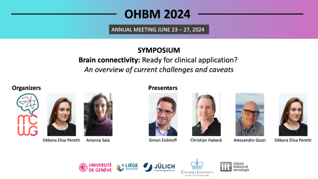
We are pleased to announce that OHBM 2024 will feature a symposium organized by members of the MCWG!

Organized by Débora Peretti and Arianna Sala, the symposium is entitled “Brain connectivity: ready for clinical application? An overview of current challenges and caveats” and will feature presentations from Simon Eickhoff, Christian Habeck, Alessandro Gozzi, and Débora Peretti. In this symposium we discuss some of the most pressing methodological considerations that still need to be addressed by neuroscientists in order to obtain robust connectivity markers for inference and prediction. Both statistical and biological considerations will be covered, on the grounds that not only prediction of phenotypes from activation/connectivity data, but also understanding the mechanisms underlying inter-individual connectivity differences, are necessary to obtain robust, generalizable topographic substrates. Finally, an overview of the translation of connectivity metrics into clinical practice will be presented. Date and time to be announced.
Members of the MCWG are co-organizing the 12th European Conference on Clinical Neuroimaging in March in Leipzig, featuring invited talks on molecular imaging, total-body PET, imaging in psychiatry and ATN concept in Alzheimer’s disease. Register now.
Please send us information about events related to brain connectivity and molecular connectivity that you wish to share with the community for consideration no later than the final day of each month using this form.
Please submit any news, including publications you would like highlighted or job listings related to brain connectivity and molecular connectivity that you would like to share with the community for consideration no later than the final day of each month using this form.
Each month, we will feature a member of the MCWG and inclide a brief Q&A!
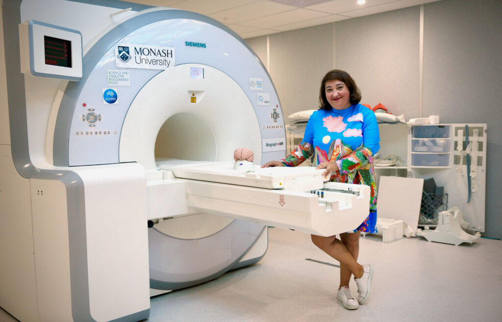
This month we highlight Dr. Sharna Jamadar, member of the MCWG Steering Committee.
Dr. Sharna Jamadar is Associate Professor (Research) and NHMRC Emerging Leader Fellow at Monash Biomedical Imaging and Turner Institute for Brain and Mental Health.Sharna’s research is focused on understanding how our life experiences change our brains, and how this may confer resilience to the ageing process. She is particularly interested in studying the neural bases of cognitive control, and has explored the consequences of disruption of cognitive control in a number of conditions, including healthy ageing, psychosis (schizophrenia, bipolar disorder) and substance abuse. She also has an emerging interest in the neuroscience of parenthood and is supervising two student projects in this theme. Sharna is an expert in multimodal neuroimaging, and uses a number of imaging techniques to understand cognitive control, including positron emission tomography (PET), functional magnetic resonance imaging (fMRI), electroencephalography (EEG) and oculomotor measures (eye-tracking). Sharna’s team leads the development of simultaneous functional MRI-PET imaging in humans at the facility. Sharna’s team has developed novel MRI-PET measures that provide high resolution mapping of the function, structure, and metabolic efficiency of the brain. For the first time, these new techniques allow researchers to study task-related changes in brain function and metabolism with a temporal resolution below 20sec. This work will have substantial implications for our understanding of how the brain dynamically uses energy; and Sharna will use these new methods to understand how the metabolic efficiency of the brain changes across the lifespan.

Dr. Sharna Jamadar has graciously responded to our feature questionnaire!
What sparked your interest in molecular imaging or led you to focus on research in molecular imaging?
I trained as a cognitive neuroscientist in EEG and fMRI, and at the time that I was doing my training (2005-2010), I (somewhat naively) thought of PET as a functional neuroimaging tool that had been superseded by fMRI. In terms of functional brain mapping and brain-cognition relationships, the vast majority of the work I was reading, and doing myself, was focused on electrophysiology and haemodynamics. From the beginning though, I always had a healthy dose of scepticism about what we can and can’t do with fMRI – you may remember a rather famous paper by Logothetis with that exact title.
In 2015 I returned to work after a year’s maternity leave, and this was a time of transition both personally and professionally. At that time, we were installing a new PET/MR scanner at our institute, and I was really excited about the possibilities this new technology was offering. In particular, I was intrigued by how FDG-PET can be considered a more direct and quantitative measure of neuronal function than fMRI. I was moving my research into the area of cognitive ageing, and this was exciting to me, as there are lots of challenges to using fMRI in older adults, particularly if we want to compare them to younger adults, which I did. I was particularly inspired by the Villien et al. 2014 paper in NeuroImage, with the proposed method for functional PET (fPET) for brain mapping. We noticed that their approach didn’t provide simultaneous task contrast for both fMRI and fPET, so our first pilot study was a simple replication and extension of that work to allow this. I’d never used PET before, let alone fPET, and this was a really fun time playing in the lab and figuring out this new method for measuring brain function. It was a great time – we tried lots of new things in the scanner, as well as with the data, collaborating across radiography, nuclear medicine technology, engineering, maths, and physics – with me the lowly neuroscientist trying to figure out what it all means!
As a follow-up to this work, I quickly shifted to considering how we might use PET to measure connectivity in the brain. The human brain mapping community had been really focused on resting connectivity for some time, and I’d done a bit of work in this area too. We’d just published a paper showing how the fMRI connectome is confounded by individual differences in haemoglobin in an older population, and so the idea that we might be able to get a less confounded measure of connectivity was really appealing. Of course, I was “scooped” back in the 1980s by Barry Horwitz and colleagues, as well as the greater PET community in the subsequent years. However, after reading the literature, I realised that the definition of connectivity used in PET was different to how I’d use it if I was looking at an EEG or fMRI measure. In other functional connectivity approaches (I use the term ‘functional connectivity’ in its broadest sense, as a measure that can be made using any functional neuroimaging approach, including EEG, fMRI, MEG, etc.), we correlate the timeseries of activity within subjects to estimate a connectome, whereas with PET the standard was an across-subject approach. So, we thought we’d try to use fPET to estimate a timeseries to correlate, in a similar way. We did this, and published what we think is the first ‘metabolic’ connectome that is estimated using within-subject methodology. The work was hard but rewarding, and we still have lots to learn about how to best process this data, and interpret it.
And it’s this interpretation that’s made me put my hand up and declare myself a diehard convert to molecular connectivity. I’ve focused exclusively on using FDG as a radiotracer, primarily because it’s easy to source, but also because of my early focus on neuronal activity. However, if you’re interested in human physiology, how can you go past the target that literally fuels the brain and body?! My group has subsequently shifted to focus on how individual differences in systemic glucose metabolic efficiency (insulin resistance) influences brain function, the metabolic connectome, and cognition. We’ve got some new results currently under review and about to be submitted that we’re really excited to share with the community in this space.
What is your role in the Molecular Connectivity Working Group and what have you been contributing to/working on with the Molecular Connectivity Working Group?
I’m involved as a Board Member, as well as a member for the symposium council. I’m excited at the opportunity to bring together international interdisciplinary experts in connectivity, to discuss how we can ‘speak the same language’ between the molecular connectivity and other functional connectivity disciplines.
In what ways do you imagine the impact of molecular connectivity will have in advancing our understanding of brain function?
I am really enthusiastic about the possibilities in this space to make important discoveries about brain function. To date, I don’t think the human brain imaging community has really capitalised on the strengths of PET and other modalities to measure molecular targets in vivo in the brain. As such, we have a narrow concept of brain function that we’ve derived from electrophysiology and haemodynamic approaches, that do not adequately take into account the range of molecular targets that we know are important for brain function. As a multimodal neuroimager, I’ve always been inspired by that famous cartoon of the man looking under the streetlamp for his keys. When a passer-by asks him where he lost his keys, he points to a place outside the light and says, ‘Over there, but the light is best over here’. We’re almost always using tools to answer questions that they’re not quite suited to answer. I like to imagine that each neuroimaging method sheds a little more light on the situation: the beams will probably overlap at some level, but also show us something new too. I’m hoping that with the development of the fPET approach, and a renewed focus on molecular connectivity, we will be able to shed new light on human brain function that we couldn’t have achieved with the other methods we have at our disposal.
One aspect that I am excited about, is the ability to use fPET and molecular connectivity with a range of radiotracers to measure dynamic brain activity. During my PhD, I was reading a lot about the role of different neurotransmitters in different cognitive processes, however, these ‘models’ were poorly specified, and had no direct measurements of the neurotransmitter targets to support them. With fPET and molecular connectivity, we can directly measure these targets and test these hypotheses. I think that these methodological advances together with the large improvements in PET detector technology means that we will be seeing a lot of new discoveries in the coming years about the molecular bases of cognition.

The MCWG is made up of four international and multidisciplinary councils dedicated to promoting molecular connectivity research via dissemination of methods, results, collaboration, and resource sharing (e.g. datasets, tools) within the scientific community. We encourage the neuroscientific community to take an integrative perspective in study of the brain connectome, where various methods including MRI-based techniques, electrophysiological tools, and molecular imaging advance our understanding of the brain. Please find fundamental questions outlined here: “Brain connectomics: time for a molecular imaging perspective?”
Our website can be found here. We also invite you to join the MCWG!