

Welcome back from what we hope was a restful, sun-filled summer break! Whether you spent the last few months traveling, spending time with family, or simply enjoying the slower pace of the season, we are thrilled to have our community back together.
Date: October 16th, 2026
Time: 12:00 UTC
Registration: Please register here.
Title: Four distinct trajectories of tau deposition identified in Alzheimer’s disease
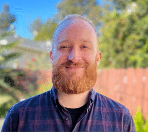
Speaker: Dr. Jake Vogel is an Assistant Professor and head of the Neurodegenerative Research Unit, a division of the Department of Clinical Science at Lund University. His lab, the Dementia Multi-Omics and Neuroimaging (DeMON) lab, focuses on integrating sophisticated statistical and AI approaches to large multi-modal data to extract novel biological insights and useful tools for neurodegenerative disease. Jake trained at UC-Berkeley, got his PhD at McGill University, and did a postdoc at the University of Pennsylvania, becoming an expert along the way in neuroimaging, aging, AI and bioinformatics. As a SciLifeLab and Wallenberg National Program Data-driven Life Sciences (DDLS) fellow, he now dedicates his time toward better understanding how neurodegenerative diseases work, and building innovative tools to improve clinical care.
Abstract: Alzheimer’s disease (AD) is characterized by the accumulation of Aβ plaques and tau tangles in the brain. While memory loss is a primary symptom of AD, clinical variants with distinct cognitive deficits are well known to the field, and patients tend to vary widely in the speed and nature of the clinical progression. In 2021, we used a large multi-site PET dataset to show that AD tau pathology tends to accumulate in four separate patterns – subtypes of AD – accompanied by distinct clinical profiles. In this talk, I will give an update on work we’ve done since to further characterize these different AD progression patterns and to better understand their origin. This work encompasses investigations into brain structural, functional, molecular (PET, genetics, proteomics) and postmortem investigations across numerous datasets.
Each month, we will feature a member of the MCWG and have a brief Q&A!
This month please enjoy our highlight of Prof Dr Joana Pereira, member of the MCWG Steering Committee.

Dr Joana B. Pereira is an Associate Professor at Karolinska Institute, where she leads the Brain Connectomics Lab. Her research interests include neuroimaging, blood and cerebrospinal fluid biomarkers, brain connectivity and machine learning. She has authored more than 120 articles and reviews, and has co-developed BRAPH, an open-access software for analyzing brain connectivity using graph theory and deep learning. She also co-authored the book “Deep Learning Crash Course” (No Starch Press, 2024), is the scientific coordinator of an European Alliance focused on neurotechnology – NeurotechEU – at KI, and the chair of the interdisciplinary conference “Emerging Topics in Artificial Intelligence”. Finally, she is the recipient of the prestigious 2021 De Leon prize for best neuroimaging article in Alzheimer’s disease.
Prof Dr Pereira has graciously responded to our feature questionnaire:
What sparked your interest in molecular imaging or led you to focus on research in molecular imaging?
My background is primarily in brain connectivity, and I became interested in molecular imaging because I felt that connectivity alone could tell us where the brain is changing, but not necessarily why. PET imaging provides a unique window into the molecular processes underlying those network changes, including metabolism and protein pathology. This led me to become particularly interested in combining molecular imaging with network approaches to understand neurodegenerative diseases at the level of individual patients.
What is your role in the Molecular Connectivity Working Group, and what have you been contributing to or working on within the group?
Within the Molecular Connectivity Working Group, I have been particularly interested in developing and applying methods to construct molecular connectivity networks at the individual level. Much of my work has focused on metabolic connectivity using amyloid and tau PET and on understanding how they change in Alzheimer’s disease and relate to cognition and disease severity. I am also interested in extending this framework across molecular imaging modalities and integrating molecular connectivity with structural and functional brain networks.
In what ways do you imagine molecular connectivity will advance our understanding of brain function?
Molecular imaging is traditionally analyzed region by region, but the brain does not function as a collection of independent regions. Molecular connectivity allows us to ask how molecular processes are coordinated across the brain and how this organization relates to structural and functional networks. I think its greatest potential is ultimately at the individual level, where molecular networks could help us understand why patients with apparently similar levels of pathology can have very different clinical trajectories.
What do you think are the most important challenges in current brain connectivity research, or which unsolved/underappreciated issues should the community address?
One major challenge is that we often treat connectivity as if there were a single “true” brain network, when different modalities capture fundamentally different biological processes. We still understand surprisingly little about how structural, functional, metabolic, and molecular networks relate to one another—and how these relationships change with aging and disease. Another important challenge is moving from group-average networks toward reliable individual-level measures that are biologically meaningful and clinically useful.
What is your favorite mentoring memory—either a story about a mentor’s impact on you or your impact on a mentee?
Some of my favorite mentoring moments are when a student starts questioning my ideas rather than simply following them. There is a point when you realize that someone you have been supervising has developed their own scientific intuition and is beginning to take the project in directions you had not considered. For me, that transition from teaching someone how to do research to actually learning from them is one of the most rewarding parts of mentoring.
What scientist or scientific achievement do you most admire?
I particularly admire Santiago Ramón y Cajal, not only for establishing the neuron doctrine but for realizing, from what were essentially static images, that the nervous system had an extraordinarily organized architecture. His work combined careful observation with an ability to infer principles of brain organization that could not yet be directly measured. In a way, modern connectomics is still pursuing the same fundamental question he was asking more than a century ago: how does the organization of individual elements give rise to a functioning brain?
Dear Colleagues,
We are delighted to announce the call for applications to the doctoral candidate position in Edinburgh for the PREFERENCE project is now open! This is a highly prestigious MSCA doctoral network coordinated by Ronald Boellaard with 15 doctoral candidates distributed across multiple institutions in Europe. The doctoral candidate in Edinburgh will work on development of new methodologies for whole-body network analysis of LAFOV PET datasets, and will be embedded into the PET is Wonderful team in Edinburgh.
Please find out more at the job post and the website: https://preference-consortium.eu/
There are brain focused project positions available: https://preference-consortium.eu/?page_id=269
The deadline for application is the 1st November 2026.
A multimodal total-body dynamic [18F]FDG PET/CT/MRI dataset of 100 healthy humans
The Multimodal-HC dataset provides comprehensive multimodal imaging data from 100 healthy participants, stratified by age and sex to capture physiological variation across the adult lifespan. This dataset addresses the critical need for normative reference data in quantitative PET imaging research.
Read the full publication in Scientific Data.
Just a reminder to the community that MCWG also has a vast list of open dataset available for anyone interested in exploring, building, or conducting research: Imaging Datasets
📝Vulnerability of locus coeruleus connections to aging and Alzheimer’s disease
In this study, Zufiria-Gerbolés and colleagues aimed at understanding the role of the locus coeruleus in coordinating communication between brain regions through its widespread connections in healthy aging and Alzheimer’s disease patients.
Read the full study in Alzheimer’s and Dementia.
📝Metabolic Connectivity Alterations in Amyotrophic Lateral Sclerosis: Individual Network Analysis Based on Wasserstein Distances
Tang and colleagues sought to introduce the Wasserstein distance as a method for constructing subject-specific metabolic connectivity metrics from static FDG PET scans, applying to healthy volunteers and amyotrophic multiple sclerosis patients and comparing it to Kullback–Leibler divergence similarity estimation.
Read the full study in Imaging Neuroscience.
📝Alterations in regional cerebral perfusion in drug-induced Parkinsonism: Spatial covariance analyses of early-phase 18F-FP-CIT PET data
In this study Chun and colleagues aimed to investigate distinct cerebral perfusion patterns on early-phase [18F]FP-CIT PET in patients with DIP.
Read the full study in Parkinsonism & Related Disorders.
Key Findings:
📝Distinct brain metabolic patterns reflect biological heterogeneity and relate to clinical outcome in chronic migraine with medication overuse
Chronic migraine with medication overuse (CMwMO) is a disabling phenotype of migraine with heterogenous responses to medication withdrawal and preventive treatments. Liu et al investigated whether baseline brain metabolic patterns reflect biological heterogeneity associated with 1-year clinical outcome in CMwMO.
Read the full study in Cephalalgia.
Key Findings:
📝Personalized metabolic connectome analysis reveals glucose heterogeneity in ischemic cerebrovascular disease
In this study, Cui and colleagues constructed individualized metabolic networks for patients with Ischemic cerebrovascular disease (ICVD) using [18F]FDG PET data. By comparing topological properties across preoperative, postoperative, and control groups, we aimed to elucidate the patterns of metabolic remodeling associated with ICVD.
Read the full study in Eur J Nucl Med Mol Imaging.
Key Findings:
The MCWG Outreach Council invites you to submit announcements or information about papers, conferences, presentations or other events or news related to brain and molecular connectivity as well as any positions available or job opportunities that you wish to publicize and share with the community!
Please submit any material for consideration by the final day of each month using this form – thank you!
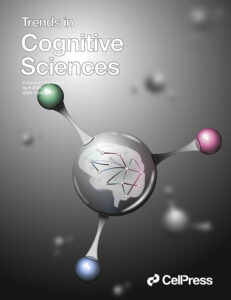
The MCWG is made up of four international and multidisciplinary councils dedicated to promoting molecular connectivity research via dissemination of methods, results, collaboration, and resource sharing (e.g. datasets, tools) within the scientific community. We encourage the neuroscientific community to take an integrative perspective in study of the brain connectome, where various methods including MRI-based techniques, electrophysiological tools, and molecular imaging advance our understanding of the brain. Please find fundamental questions outlined here: “Brain connectomics: time for a molecular imaging perspective?”
Our website can be found here. We also invite you to join the MCWG!