
In the spirit of Halloween and the mysteries that lie beyond what is immediately visible, we are reminded of the many molecular mechanisms underlying brain connectivity that remain to be fully understood. Through collaborative efforts within the MCWG community, we continue to advance our understanding of the molecular basis of brain connectivity and its role in brain function.

Congratulations to Tommaso Volpi, MD, PhD, Associate Research Scientist in Radiology and Biomedical Imaging, on being named to the 2026 “Ones to Watch” list by the Society of Nuclear Medicine and Molecular Imaging (SNMMI)!
The SNMMI “Ones to Watch” list recognizes early-career professionals with the potential to shape the future of precision medicine across the field. This recognition highlights Dr. Volpi’s contributions to nuclear medicine and molecular imaging, particularly his innovative work using mathematical modeling of PET data to investigate brain physiology and better understand brain disorders. His research includes applications to neurological and psychiatric conditions such as Schizophrenia and Parkinson’s disease, with the goal of extracting new insights from PET imaging to advance our understanding of brain function and disease.
We are proud to celebrate Dr. Volpi’s recognition and his contributions to this rapidly evolving field. Please join us in congratulating Tommaso on this well-deserved honor!
Date: October 16th, 2026
Time: 12:00 UTC
Registration: Please register here.
Title: Four distinct trajectories of tau deposition identified in Alzheimer’s disease
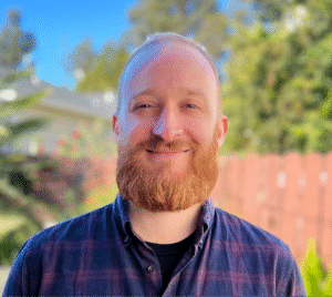
Speaker: Dr. Jake Vogel is an Assistant Professor and head of the Neurodegenerative Research Unit, a division of the Department of Clinical Science at Lund University. His lab, the Dementia Multi-Omics and Neuroimaging (DeMON) lab, focuses on integrating sophisticated statistical and AI approaches to large multi-modal data to extract novel biological insights and useful tools for neurodegenerative disease. Jake trained at UC-Berkeley, got his PhD at McGill University, and did a postdoc at the University of Pennsylvania, becoming an expert along the way in neuroimaging, aging, AI and bioinformatics. As a SciLifeLab and Wallenberg National Program Data-driven Life Sciences (DDLS) fellow, he now dedicates his time toward better understanding how neurodegenerative diseases work, and building innovative tools to improve clinical care.
Abstract: Alzheimer’s disease (AD) is characterized by the accumulation of Aβ plaques and tau tangles in the brain. While memory loss is a primary symptom of AD, clinical variants with distinct cognitive deficits are well known to the field, and patients tend to vary widely in the speed and nature of the clinical progression. In 2021, we used a large multi-site PET dataset to show that AD tau pathology tends to accumulate in four separate patterns – subtypes of AD – accompanied by distinct clinical profiles. In this talk, I will give an update on work we’ve done since to further characterize these different AD progression patterns and to better understand their origin. This work encompasses investigations into brain structural, functional, molecular (PET, genetics, proteomics) and postmortem investigations across numerous datasets.
Each month, we will feature a member of the MCWG and have a brief Q&A!
This month please enjoy our highlight of Valentina Turmacu, a member of the MCWG.
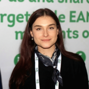
Valentina Turmacu is a Doctoral Researcher in the Nuclear Medicine and Molecular Imaging group at KU Leuven. Her PhD focuses on optimising ultra-high-resolution dynamic FDG brain PET on the NeuroEXPLORER scanner, for absolute metabolic quantification and functional activation mapping. She holds a BSc and an MSc in Biomedical Engineering (Medical Imaging) from the University of Groningen. Her bachelor’s thesis examined how PET reconstruction methods affect kinetic modelling in dynamic preclinical PET, and the protocols she optimised were later adopted into the University Medical Centre Groningen (UMCG) department’s standard workflow. As a research assistant at UMCG, she went on to evaluate image-derived input functions for quantifying P-glycoprotein function with [¹⁸F]MC225 PET. During her master’s, she compared short and long axial field-of-view PET for brain imaging. She then completed her master’s thesis at Positrigo AG, working on advanced reconstruction methods for a dedicated brain PET scanner.
Ms Turmacu has graciously responded to our feature questionnaire:
What sparked your interest in molecular imaging or led you to focus on research in molecular imaging?
I have always been drawn to physics, and molecular imaging felt like the place where physics becomes directly meaningful for medicine. Most imaging shows us what the body looks like, while PET lets us measure what it is actually doing, from glucose metabolism to transporter function. During my bachelor’s thesis in Groningen, which focused on PET reconstruction methods, I was struck by how much the choices made before an image is even seen can shape the biological conclusions drawn from it. That realisation has guided my work ever since: I want to make sure the numbers we extract from PET images are ones we can truly trust.
In what ways do you imagine molecular connectivity will advance our understanding of brain function?
Molecular connectivity adds a biological layer to our picture of brain networks. MRI-based connectivity tells us how regions are wired or how their activity co-fluctuates. Molecular connectivity can reveal how processes such as metabolism, receptor availability or protein pathology are coordinated across the brain. I am particularly excited about what new scanner technology will make possible. Long axial field-of-view systems and dedicated brain PET scanners offer much higher sensitivity and resolution, making dynamic, low-dose and repeated imaging far more feasible. Over time, I hope this will allow us to build reliable molecular networks for individual subjects and follow how they change. We could even look beyond the brain itself, at how brain networks interact with the rest of the body.
What do you think are the most important challenges in current brain connectivity research, or which unsolved/underappreciated issues should the community address?
In my view, an underappreciated issue is how strongly molecular connectivity measures depend on the imaging chain behind them. Reconstruction algorithms, resolution, partial-volume effects, noise and scanner differences all influence regional PET values. Because connectivity is built from the relationships between those values, these effects can shape the resulting networks in ways that are easy to mistake for biology. As the field moves towards individual-level and multi-centre analyses, we need more systematic work on harmonisation and a clearer understanding of how robust network measures are to acquisition and processing choices. I’m also looking forward to more dynamic PET in connectivity research. Its kinetic information can yield more specific and physiologically interpretable networks than static uptake alone, provided we can quantify it reliably and, ideally, non-invasively.
MCWG have added a new list of open datasets with others already available for anyone interested in exploring, building, or conducting research: Imaging Datasets
Check out the new adds on:
Van Laere, K., Koole, M., & De Hoon, J. (2025). Leuven PET UCB-J dataset 1 in control participants Data [Dataset]. In PublicNeuro Datasets. Neurobiology Research Unit, Copenhagen University Hospital Rigshospitalet. https://doi.org/10.70883/bkwa8149
Van Laere, K., & Van Aalst, J. (2025). Leuven PET UCB-J dataset 2 in control participants Data [Dataset]. In PublicNeuro Datasets. Neurobiology Research Unit, Copenhagen University Hospital Rigshospitalet. https://doi.org/10.70883/fiyq9929
Vanhaute, H., Michiels, L., Vanderlinden, G., & Vandenbulcke, M. (2025). Leuven PET UCB-J dataset 3 in control participants Data [Dataset]. In PublicNeuro Datasets. Neurobiology Research Unit, Copenhagen University Hospital Rigshospitalet. https://doi.org/10.70883/guma1819
Van Laere, K., Delva, A., & Vandenberghe, W. (2025). Leuven PET UCB-J dataset 4 in control participants Data [Dataset]. In PublicNeuro Datasets. Neurobiology Research Unit, Copenhagen University Hospital Rigshospitalet. https://doi.org/10.70883/miiq4096
Van Laere, K., Delva, A., & Vandenberghe, W. (2025). Leuven PET UCB-J dataset 5 in control participants Data [Dataset]. In PublicNeuro Datasets. Neurobiology Research Unit, Copenhagen University Hospital Rigshospitalet. https://doi.org/10.70883/wsyq4663
Multimodal neuroimaging dataset UCB-J project including T1-weighted MRI, [11C]UCB-J PET, and FDG PET. The dataset includes healthy controls (young and older) and patients with Parkinson’s disease spectrum disorders (PD, DLB, PDD), organized in BIDS format. In PublicNeuro Datasets. Aarhus University Katrine B. Andersen, Per Borghammer. Link
Multimodal total-body 18F-FDG PET-CT-MRI – static images. Christian Hinge, Frederikke Engel Høi-Hansen, Rasmus Hvass Hansen, Kirsten Korsholm, Helle Hjorth Johannesen, Elisabeth Albrech-Beste, Eunice Sánchez Saxtoft, Vibeke Jensen, Christina Schulze, Christina Mikkelsen, Cyril Pernet, Janna Nissen, Sisse Rye Ostrowski, Barbara Malene Fischer, Claes Nøhr Ladefoged, Flemming Littrup Andersen. In PublicNeuro Datasets. Link
multimodal total-body dynamic 18F-FDG PET-CT-MRI. Christian Hinge, Frederikke Engel Høi-Hansen, Rasmus Hvass Hansen, Kirsten Korsholm, Helle Hjorth Johannesen, Elisabeth Albrech-Beste, Eunice Sánchez Saxtoft, Vibeke Jensen, Christina Schulze, Christina Mikkelsen, Cyril Pernet, Janna Nissen, Sisse Rye Ostrowski, Barbara Malene Fischer, Claes Nøhr Ladefoged, Flemming Littrup Andersen. In PublicNeuro Datasets. Link
Hinge, C., Engel Høi-Hansen, F., Hvass Hansen, R., Korsholm, K., Hjorth Johannesen, H., Albrech-Beste, E., Sánchez Saxtoft, E., Jensen, V., Schulze, C., Mikkelsen, C., Pernet, C., Nissen, J., Rye Ostrowski, S., Malene Fischer, B., Nøhr Ladefoged, C., & Littrup Andersen, F. (2025). multimodal total-body source data 18F-FDG PET-CT-MRI Data [Dataset]. In PublicNeuro Datasets. Neurobiology Research Unit, Copenhagen University Hospital Rigshospitalet. https://doi.org/10.70883/jzjh3431
M. Knudsen, G., & G. Frokjaer, V. (2026). Serotonine transporter data in healthy and obese participants. Data [Dataset]. In PublicNeuro Datasets. Neurobiology Research Unit, Copenhagen University Hospital Rigshospitalet. https://doi.org/10.70883/gbwn3773
M. Fisher, P., K. Madsen, M., & M. Knudsen, G. (2026). Serotonine transporter PET data in healthy controls during bright-light therapy Data [Dataset]. In PublicNeuro Datasets. Neurobiology Research Unit, Copenhagen University Hospital Rigshospitalet. https://doi.org/10.70883/jeka8722
Erritzoe, D., G. Frokjaer, V., & K. Holst, K. (2026). Serotonine transporter imaging in MDMA or hallucinogenic drug users and non using controls Data [Dataset]. In PublicNeuro Datasets. Neurobiology Research Unit, Copenhagen University Hospital Rigshospitalet. https://doi.org/10.70883/zkcf9211
Mc Mahon, B., B. Andersen, S., K. Holst, K., G. Frokjaer, V., & M. Knudsen, G. (2026). Serotonine transporter imaging in seasonal affective disorder Data [Dataset]. In (Editor), PublicNeuro Datasets. Neurobiology Research Unit, Copenhagen University Hospital Rigshospitalet. https://doi.org/10.70883/frbp6839
Haugbøl, S., H. Pinborg, L., & M. Knudsen, G. (2026). Serotonergic brain architecture of early Alzheimers Disease. Data [Dataset]. In (Editor), PublicNeuro Datasets. Neurobiology Research Unit, Copenhagen University Hospital Rigshospitalet. https://doi.org/10.70883/mrpf9876
G. Frokjaer, V., & M. Knudsen, G. (2026). Familial risk for affective disorders and serotonergic brain architecture Data [Dataset]. In (Editor), PublicNeuro Datasets. Neurobiology Research Unit, Copenhagen University Hospital Rigshospitalet. https://doi.org/10.70883/neot1858
Dear Colleagues,
We are delighted to announce the call for applications to the doctoral candidate position in Edinburgh for the PREFERENCE project is now open! This is a highly prestigious MSCA doctoral network coordinated by Ronald Boellaard with 15 doctoral candidates distributed across multiple institutions in Europe. The doctoral candidate in Edinburgh will work on development of new methodologies for whole-body network analysis of LAFOV PET datasets, and will be embedded into the PET is Wonderful team in Edinburgh.
Please find out more at the job post and the website: https://preference-consortium.eu/
There are brain focused project positions available: https://preference-consortium.eu/?page_id=269
The deadline for application is the 1st November 2026.
📝Left Cerebellar-Right Frontal Uncoupling in Female First-Episode Major Depressive Disorder: Synaptic Evidence from [18F]UCB-H PET
This study by Wang and colleagues aimed at assessing the connections between cerebellum and cortex using the synaptic density radiotracer [18F]UCB-H in a cohort composed of women with first-episode major depressive disorder.
Read the full study in CNS Neuroscience & Therapeutics.
Key Findings:
📝Metabolic networks on PET-based C-Atlas for diagnosis of Alzheimer’s disease
Ahn and colleagues proposed the construction of a metabolic network using a new FDG-PET based atlas, the Coefficient-Atlas (C-Atlas), with nodes defined through kernel density estimation, in order to enhance Alzheimer’s disease classification.
Read the full study in IEEE Xplore.
Key Findings:
📝Directed information flow across the metabolic network of the human brain
In this work by Deery and colleagues, Granger causality analysis is used as a modelling framework for directed metabolic connectivity using the functional FDG-PET scanning approach. They analysed specific networks and their associations with cognition and psychosocial function.
Read the full study in Network Neuroscience.
Key Findings:
📝Associations of visceral adipose tissue metabolism with DMN-centered cross-network metabolic expression concordance and cognitive impairment in Alzheimer’s disease
Xia and colleagues investigated the associations between glucose metabolism in visceral adipose tissue and subcutaneous adipose tissue with abnormalities in large-scale brain metabolic networks and cognitive impairment in Alzheimer’s disease patients.
Read the full study in European Journal of Nuclear Medicine and Molecular Imaging.
Key Findings:
📝A Comparison of Brain Metabolic Connectivity Methods: Topology, Cognition, Age, Structure, and Genetics
In this study, Deery and colleagues compared metabolic connectivity approaches in 80 participants who underwent simultaneous dynamic PET with 18F-fluorodeoxyglucose and functional magnetic resonance imaging (fMRI) scans and cognitive testing. Six connectivity approaches were compared, and three approaches that use distance-based measures to estimate individual-level connectivity. fMRI connectivity was also calculated.
Read the full study in bioRxiv.
Key Findings:
The MCWG Outreach Council invites you to submit announcements or information about papers, conferences, presentations or other events or news related to brain and molecular connectivity as well as any positions available or job opportunities that you wish to publicize and share with the community!
Please submit any material for consideration by the final day of each month using this form – thank you!
The MCWG is made up of four international and multidisciplinary councils dedicated to promoting molecular connectivity research via dissemination of methods, results, collaboration, and resource sharing (e.g. datasets, tools) within the scientific community. We encourage the neuroscientific community to take an integrative perspective in study of the brain connectome, where various methods including MRI-based techniques, electrophysiological tools, and molecular imaging advance our understanding of the brain. Please find fundamental questions outlined here: “Brain connectomics: time for a molecular imaging perspective?”
Our website can be found here. We also invite you to join the MCWG!